Ectopic Pancreatic Tissue - A Rare Case Of Subserosal Tumor In The Jejunum [Case Report]
In 75% of the cases, the ectopic pancreatic tissue is in the submucosa, with around 13% of these tumors in the subserosa. This is interesting because of the tumor’s location.
Author:Suleman ShahReviewer:Han JuSep 23, 202489.1K Shares1.4M Views

Ectopic pancreas is defined as a pancreatic tissue in an abnormal location with no ductal, anatomical, neuronal, vascular communication with the main body of the pancreas.
The incidence of heterotopic pancreas in autopsy studies is approximately 0.55% to 13.7% and as low as 0.2% in laparotomies.
Most frequently, it is located in the stomach, the duodenum, the proximal jejunum or the Meckel's diverticulum.
Rarely, it is seen in the ileum, the gallbladder, the bile ducts, the splenic hilum, the umbilicus and the liver.
The present report describes a rare case where the ectopic pancreatic tissue was located in the jejunum as a subserosal tumor.
Case Report
A 53-year-old woman presented in our hospital for an elective sigmoidectomy (or sigmoid colectomy) due to chronic diverticulitis.
The physical examination did not reveal any abnormal findings.
Laboratory exams were normal:
- WBC (white blood count) of 8.4 × 10³/μL (reference range: 5 to 10 × 10³/μL)
- neutrophils of 62% (reference range: 40 to 80%)
- a C-reactive protein equal to 2.6 mg/L (reference range: less than 5 mg/L)
- ESR (erythrocyte sedimentation rate) equal to 24 mm/h (reference range: 20 to 30 mm/h)
As well, all other laboratory values, including amylase and lipase, were within the reference limits.
The preoperative ultrasonographystudy of the abdomen, the endoscopic examination via colonoscopy, as well as the abdominal CT examination (with intravenous contrast) did not detect the presence of any intra-abdominal tumor.
Intraoperatively, the chronic inflamed diverticula of the sigmoid were recognized and the sigmoidectomy was performed without complications.
After the sigmoid resection, we proceeded with a macroscopic examination of the small bowel.
Incidentally, at the distal jejunum, a 2 × 3 cm yellow-white nodule was recognized.
The nodule was located in the subserosa of the jejunum and was excised. The site of incision was repaired with sutures.
The patient remained asymptomatic and without complications post-operatively, with normal lab values, and was discharged on the fifth post-operative day.
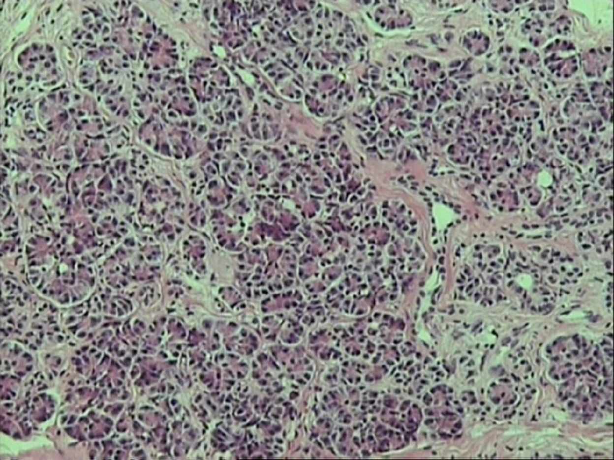
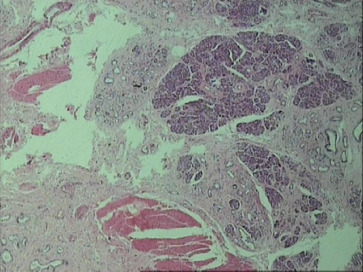
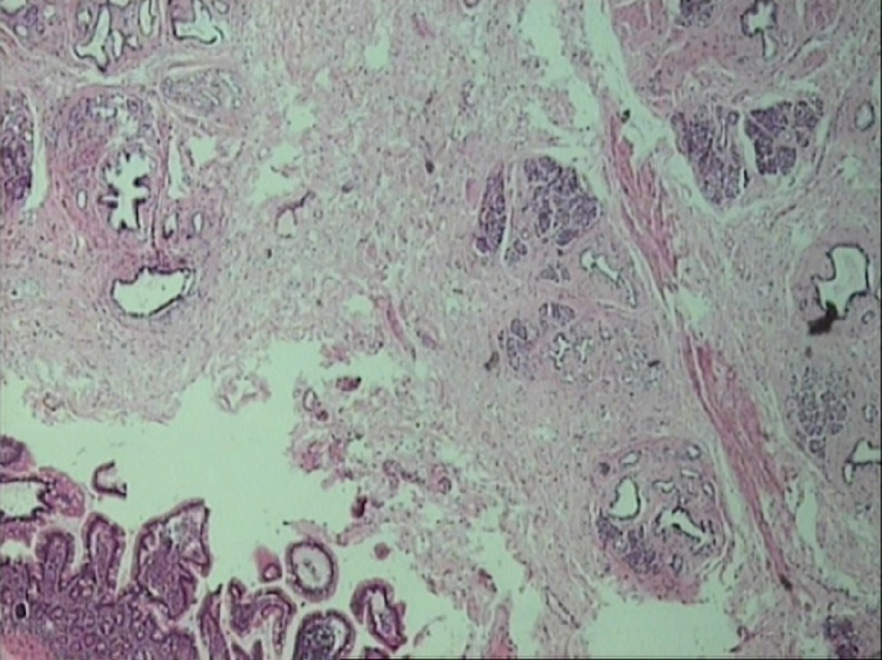
The pathology report of the nodule normal jejunal mucosa confirmed the presence of ectopic pancreatic tissue with glandular acini(salivary glands) within the muscularis propriaand subserosa of jejunum.

Discussion
The ectopic pancreatic tissue can be present anywhere along the gastrointestinal tract, and in the majority of cases it is located in the submucosa.
Heterotopic pancreas can be found in all age groups. However, it seems to be more frequent in men than in women.
Most of the patients with ectopic pancreatic tissue remain asymptomatic, and the heterotopic tissue is found incidentally at a histological exam.
Nevertheless, there are reports of patients that presented with symptoms like:
- abdominal pain
- hematemesis
- vomiting
- weight loss
- intestinal obstruction
Regarding the etiology of pancreatic heterotopia, two theories exist:
a. First theory
The first one suggests that during embryological development, buds of embryonic tissue penetrate the wall of the growing gut, separating from the main pancreas
b. Second theory
The second theory refers to an inappropriate expression of pluripotent embryonic mesenchymal tissue of the gastrointestinal tract, leading to pancreatic metaplasia.
It is commonly thought that ectopic pancreatic tissue in stomach and duodenum is a derivative of the dorsal pancreatic bud, while that in jejunum and ileum originates from the ventral one.
Histologically, most of the tumors are situated in the submucosa, rarely in the muscularis propria(muscular layer), and only seldom (around 13.5%) in the subserosal.
Despite the advances in imaging and other diagnostic tools, a preoperative diagnosis is often difficult.
Apart from thorough clinical exam, the following may be required to exclude other pathologies with similar features:
- upper GI endoscopy
- abdominal ultrasound
- CT scan
However, in cases of ectopic pancreatic tissue findings are not specific.
A treatment is required only in symptomatic patients, and in most cases, it consists of simple surgical resection. The type and extent of the resection depends on the location and the size of the lesion.
Conclusion
The preoperative diagnosis of an ectopic pancreatic tissue along the gastrointestinal tract may be difficult, due to the non-specific imaging findings.
The clinical symptoms, if present, may mimic other pathologies of the gastrointestinal tract.
Despite the fact that heterotopia of the pancreas remains rare, it should be considered in the differential diagnosis of unspecific abdominal pain and intramural gastrointestinal obstruction.
Considering the intramural location, most of the findings are located in the submucosa (75%), with only 13.5% of them in the subserosa.
In the end, more research should be carried out about ectopic pancreatic tissue.

Suleman Shah
Author
Suleman Shah is a researcher and freelance writer. As a researcher, he has worked with MNS University of Agriculture, Multan (Pakistan) and Texas A & M University (USA). He regularly writes science articles and blogs for science news website immersse.com and open access publishers OA Publishing London and Scientific Times. He loves to keep himself updated on scientific developments and convert these developments into everyday language to update the readers about the developments in the scientific era. His primary research focus is Plant sciences, and he contributed to this field by publishing his research in scientific journals and presenting his work at many Conferences.
Shah graduated from the University of Agriculture Faisalabad (Pakistan) and started his professional carrier with Jaffer Agro Services and later with the Agriculture Department of the Government of Pakistan. His research interest compelled and attracted him to proceed with his carrier in Plant sciences research. So, he started his Ph.D. in Soil Science at MNS University of Agriculture Multan (Pakistan). Later, he started working as a visiting scholar with Texas A&M University (USA).
Shah’s experience with big Open Excess publishers like Springers, Frontiers, MDPI, etc., testified to his belief in Open Access as a barrier-removing mechanism between researchers and the readers of their research. Shah believes that Open Access is revolutionizing the publication process and benefitting research in all fields.

Han Ju
Reviewer
Hello! I'm Han Ju, the heart behind World Wide Journals. My life is a unique tapestry woven from the threads of news, spirituality, and science, enriched by melodies from my guitar. Raised amidst tales of the ancient and the arcane, I developed a keen eye for the stories that truly matter. Through my work, I seek to bridge the seen with the unseen, marrying the rigor of science with the depth of spirituality.
Each article at World Wide Journals is a piece of this ongoing quest, blending analysis with personal reflection. Whether exploring quantum frontiers or strumming chords under the stars, my aim is to inspire and provoke thought, inviting you into a world where every discovery is a note in the grand symphony of existence.
Welcome aboard this journey of insight and exploration, where curiosity leads and music guides.
Latest Articles
Popular Articles
