Tension Pneumothorax - When Should You See A Doctor?
Tension pneumothorax is a life-threatening condition that happens when air keeps getting into the pleural space and getting stuck there. This puts pressure on the lungs, heart, blood vessels, and other parts of the chest, which can be fatal. The Mayo Clinic, an academic medical center in the United States that is not for profit, said that this disease is marked by a collapsed lung.
Author:Suleman ShahReviewer:Han JuOct 07, 20222.5K Shares423.3K Views

Tension pneumothoraxis a life-threatening condition that happens when air keeps getting into the pleural space and getting stuck there. This puts pressure on the lungs, heart, blood vessels, and other parts of the chest, which can be fatal. The Mayo Clinic, an academic medical center in the United States that is not for profit, said that this disease is marked by a collapsed lung.
It is life-threatening because the air pushes against the outside of your lung, causing it to collapse. A pneumothorax can be a complete lung collapse or a partial lung collapse.
Causes Of Tension Pneumothorax
Any pneumothorax has the potential to progress into a tension pneumothorax. But it is usually seen in people who have been hurt in the chest or who are getting mechanical ventilation.
Chest Injury
Injuries to the chest can happen anywhere between the neck and the stomach. The ribs, sternum, skin, fat, and muscles that protect your lungs may be damaged, as well as any of the organs inside your chest (for example, the heart or lungs). Injuries to the chest, whether blunt or penetrating, can lead to collapsed lungs. Accidental needle sticks to the chest can happen during any number of medical procedures, not just those involving the insertion of a needle during an assault or auto accident.
Lung Disease
A collapse of the lungs is more likely to occur in damaged tissue. Numerous diseases, such as cystic fibrosis, lung cancer, and pneumonia, can damage the lungs and lead to breathing difficulties. Pneumothorax is caused by the rupture of round, thin-walled air sacs in the lungs, which are a symptom of cystic lung diseases like lymphangioleiomyomatosis and Birt-Hogg-Dube syndrome.
Ruptured Air Blisters
Blebs, or small air blisters, can form on the surface of the lungs. Sometimes, these air blisters will burst, allowing air to seep into the pleural space. Air pressure changes, such as those experienced during scuba diving or a trip to a high altitude, can cause damage to air blisters. Collapsed lung disease is more common in tall, slender, and smoker people.
Symptoms And Signs
The following are the signs and symptoms that you have the collapsed lungs mentioned a while ago.
- Severe shortness of breath.
- Breathing is shallow.
- Acute chest pain.
- Low blood oxygen levels.
- Increased heart rate.
- Low blood pressure.
- Mental state change.
In severe cases, you will experience the following signs:
- Blue skin discoloration caused by low blood oxygen levels.
- Neck veins are swollen due to collapsed blood vessels that should return blood to the heart.
- The air in the pleural space can sometimes leak out and get stuck in nearby places, like the subcutaneous tissue, causing subcutaneous emphysema, or cavities, like the mediastinum, causing pneumomediastinum.
Tension Pneumothorax Treatment
Needle Thoracostomy
Tension pneumothorax is an emergency medical situation that needs treatment right away. Needle thoracostomy, which is another name for needle decompression of the chest, is used to release the air that has been trapped in the pleural space. During this process, an emergency technician or trained doctor will put a large needle through the chest wall, between the ribs, and into the pleural space.
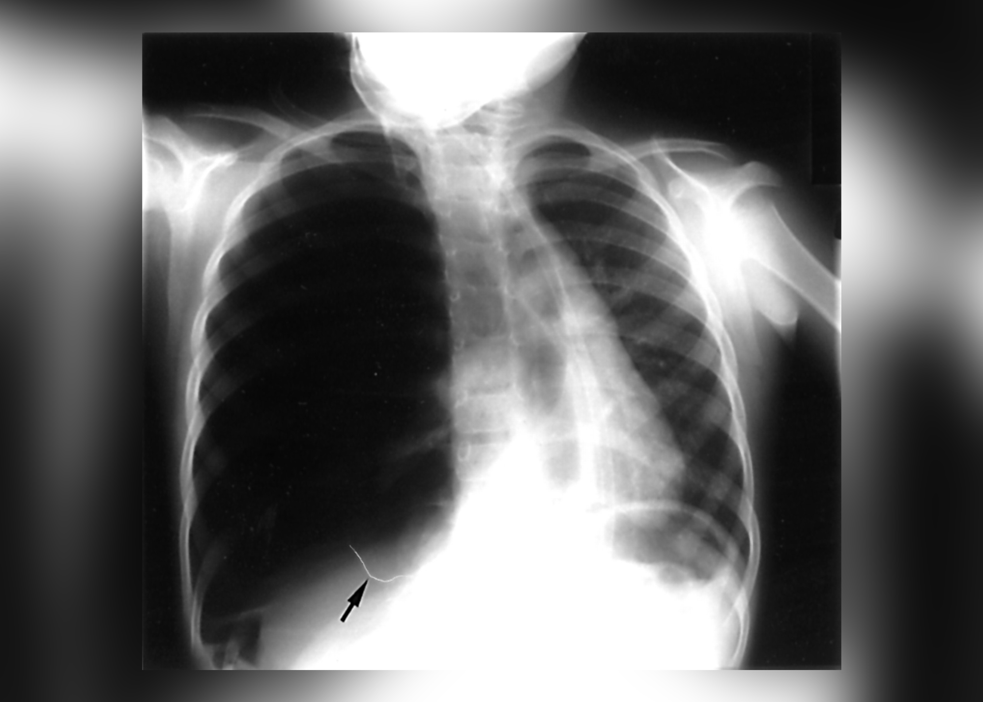
This procedure can save a person's life, especially before they get to the hospital, where getting to the hospital can slow down treatment. Then, after decompressing the needle, the needle is left in place until a more permanent chest tube can be put in to help release the remaining air. After the chest tube is put in, a chest X-ray is usually done to see where the tube is and if the lung has fully expanded again.
People Also Ask
What Happens In A Tension Pneumothorax?
Tension pneumothorax happens when air builds up between the chest wall and the lung. This raises the pressure in the chest and makes less blood go back to the heart. Some of the symptoms are chest pain, shortness of breath, fast breathing, a racing heart, and then shock.
What's The Difference Between Pneumothorax And Tension Pneumothorax?
Pneumothorax (air in the pleural cavity) can be open (external wound) or closed (internal wound). Pleural pressure reaches equilibrium with atmospheric pressure, causing lung collapse. Tension pneumothorax occurs when air enters the chest without being expelled.
What Lung Sounds Are Heard With Pneumothorax?
Tension pneumothorax can be found when breath sounds aren't the same on both sides (or aren't there on the side with the pneumothorax), the trachea moves away from the side with the pneumothorax, neck veins get bigger, or there are signs of trouble breathing.
How Do Hospitals Remove Fluid From Lungs?
Thoracentesis is a technique used to remove air or fluid from the area around the lungs. The pleural space is reached by inserting a needle through the chest wall. The pleural space is the tiny opening between the inner chest wall and the pleura of the lung. A double layer of membranes called the pleura encircles the lungs.
Final Thoughts
Complications may arise for a variety of reasons, some of which are related to the size and severity of the pneumothorax, its underlying cause, and its treatment. Pneumothorax can reoccur or continue leaking air if the lung's opening doesn't seal completely. Pneumothorax symptoms may indicate a serious medical condition; consult a doctor if you notice any of these symptoms. Seek immediate emergency care if chest pain or breathing difficulties persist.

Suleman Shah
Author
Suleman Shah is a researcher and freelance writer. As a researcher, he has worked with MNS University of Agriculture, Multan (Pakistan) and Texas A & M University (USA). He regularly writes science articles and blogs for science news website immersse.com and open access publishers OA Publishing London and Scientific Times. He loves to keep himself updated on scientific developments and convert these developments into everyday language to update the readers about the developments in the scientific era. His primary research focus is Plant sciences, and he contributed to this field by publishing his research in scientific journals and presenting his work at many Conferences.
Shah graduated from the University of Agriculture Faisalabad (Pakistan) and started his professional carrier with Jaffer Agro Services and later with the Agriculture Department of the Government of Pakistan. His research interest compelled and attracted him to proceed with his carrier in Plant sciences research. So, he started his Ph.D. in Soil Science at MNS University of Agriculture Multan (Pakistan). Later, he started working as a visiting scholar with Texas A&M University (USA).
Shah’s experience with big Open Excess publishers like Springers, Frontiers, MDPI, etc., testified to his belief in Open Access as a barrier-removing mechanism between researchers and the readers of their research. Shah believes that Open Access is revolutionizing the publication process and benefitting research in all fields.

Han Ju
Reviewer
Hello! I'm Han Ju, the heart behind World Wide Journals. My life is a unique tapestry woven from the threads of news, spirituality, and science, enriched by melodies from my guitar. Raised amidst tales of the ancient and the arcane, I developed a keen eye for the stories that truly matter. Through my work, I seek to bridge the seen with the unseen, marrying the rigor of science with the depth of spirituality.
Each article at World Wide Journals is a piece of this ongoing quest, blending analysis with personal reflection. Whether exploring quantum frontiers or strumming chords under the stars, my aim is to inspire and provoke thought, inviting you into a world where every discovery is a note in the grand symphony of existence.
Welcome aboard this journey of insight and exploration, where curiosity leads and music guides.
Latest Articles
Popular Articles